A Central Nervous System Disease
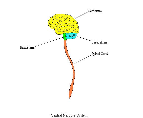
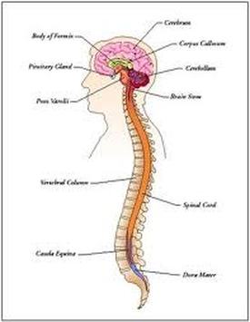
Stargardt's Disease is a neurodegenerative disease of the central nervous system. The central nervous system (CNS) is one of the two major subdivisions of the nervous system. The CNS includes the brain, brain stem, and spinal cord, which together comprise the body's main control and data center. Together with the peripheral nervous system (PNS), the CNS performs fundamental functions that contribute to an organism's life and behavior.
The nervous system has three main functions: gathering, synthesizing, and responding to stimuli. The CNS is mainly devoted to the information synthesizing function. During this step in the process, the brain and spinal cord decide on an appropriate motor output, which is computed based on a specific sensory input. The CNS regulates organ function, higher thought, and movement of the body. Thus, the CNS is commonly thought of as the control center of the body.
The Central Nervous System interacts with other systems of the body in that it directs all other systems of the body. The nervous system allows organisms to sense, organize, and react to information in the environment. The basic unit of the nervous system is the neuron, which is a highly specialized cell that communicates with the body through electrical and chemical processes. Synapses form between the neurons, allowing them to communicate to other neurons or other systems in the body
CNS directs the cardiovascular system by regulating heart and blood pressure and causing the Endothelial cells to maintain the blood-brain barrier. It directs the muscular system by allowing receptors in muscles to provide the brain with information about body position and movement while the brain controls the contraction of skeletal muscles. The brain directs the respiratory system by monitoring respiratory volume and blood gas levels and respiratory rate.
Although the brain directs these systems, other systems of the body, the Integumentary, or skin system, the endocrine system, the reproductive system, and the digestive system are all depended on and worked with very closely by the central nervous system. In the skin system, receptors in skin send sensory information to the brain and the autonomic nervous system regulates peripheral blood flow and sweat glands. In the endocrine system, hormones provide feedback to the brain to affect neural processing while Reproductive hormones affect the development of the nervous system and the hypothalamus controls the pituitary gland and other endocrine glands. In the reproductive system, reproductive hormones affect brain development and sexual behavior and the brain controls mating behavior. In the digestive system, digestive processes provide the building blocks for some neurotransmitters while the autonomic nervous system controls the tone of the digestive tract. For example, the brain controls drinking and feeding behavior and muscles for eating and elimination of waste.
|
|
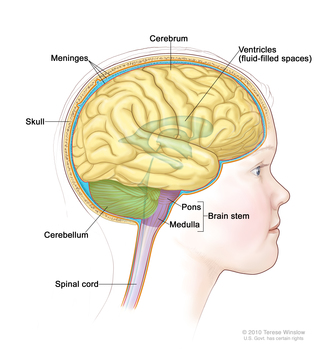
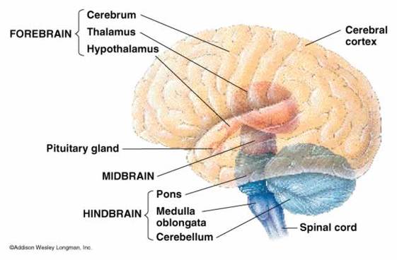
The brain is the most complex part of the human body and is an organ that serves as the center of the nervous system in all vertebrates and most invertebrate animals. The brain can be divided into three basic units; the forebrain, midbrain, and hindbrain. These seperate sections are responsible for different functions of the brain. However, they all work together as one unit. The forebrain is responsible for motor control, relaying sensory information, and controlling autonomic functions. The midbrain is involved in auditory and visual responses, as well as motor function. The midbrain connects the forebrain and hindbrain. The midbrain and hindbrain together form the brain stem. The hindbrain extends from the spinal cord and is composed of the metencephalon and the myelencephalon. Regions of the metencephalon are responsible for balance and equilibrium, movement, coordination, and the conduction of sensory information. The Medulla Oblongata which is a part of the myelencephalon is responsible for controlling autonomic functions such as breathing, heart rate, and digestion. There are many more parts within these sections of the brain which are specialized to be responsible for different specific functions of the body, he brain acts as a control center by receiving, interpreting, and directing sensory information throughout the body.
Stargardt's disease may seem like a disease of the sensory organs. However, this is not true. Stargardt's disease is a disease of the retina. The retina is the third and inner coat of the eye which is a light-sensitive layer of tissue. When vertebrates are still growing as embryos, the retina and the optic nerve originate as outgrowths of the developing brain. The retinas of the eye are actually brain tissue that has grown out from the brain. This means that the the retina is considered to be part of the central nervous system. This is why Stargardt's Disease is actually considered to be a neurodegenerative disease.
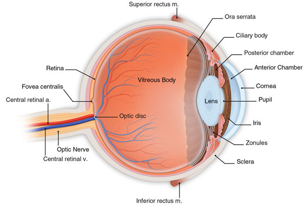
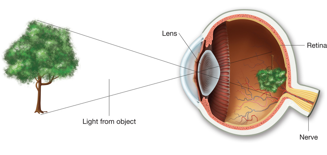
The collective function of the nonretinal parts of the eye is to keep a focused, clear image of the outside world anchored on the two retinas. Each eye is positioned in its socket by the six small extra ocular muscles. There are six for each eye; they consist of three pairs, with the muscles in each pair working in opposition, so as to take care of movements in one of three perpendicular planes. For both eyes, the job of tracking an object has to be done with a precision of a few minutes or else humans would see double. The cornea and lens together form the equivalent of the camera lens. About two-thirds of the bending of light necessary for focusing takes place at the air-cornea interface, where the light enters the eye. The lens of the eye supplies the remaining third of the focusing power, but its main job is to make the necessary adjustments to focus on objects at various distances. To focus a camera you change the distance between lens and film; we focus our eye not by changing the distance between lens and retina but by changing the shape of the rubbery, jellylike lens—by pulling or relaxing the tendons that hold it at its margin. This is so that the eye goes from a more spherical shape for near objects to flatter for far ones. A set of radial muscles called ciliary muscles produces these changes in shape.
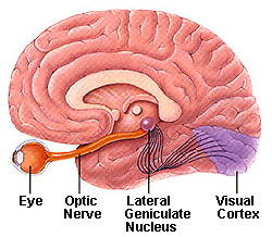
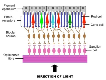
All this intricate structure exists in the interests of the retina, itself an amazing structure. It translates light into nerve signals, allows us to see under conditions that range from starlight to sunlight, discriminates wavelength so that we can see colors, and provides a precision sufficient for us to detect a human hair or speck of dust a few yards away. The retina is part of the brain, having been isolated from it early in development but having kept its connections with the brain proper through a bundle of fibers known as the optic nerve. Like many other structures in the central nervous system, the retina has the shape of a plate, in this case one about a quarter millimeter thick. It consists of three layers of nerve-cell bodies separated by two layers containing synapses made by the axons and dendrites of these cells. The tier of cells at the back of the retina contains the light receptors, the rods and cones. Rods, which are far more numerous than cones, are responsible for our vision in dim light and are out of commission in bright light. Cones do not respond to dim light but are responsible for our ability to see fine detail and for our color vision.The numbers of rods and cones vary over the surface of the retina.
In the very center, where our fine-detail vision is best, we have only cones.This rod-free area is called thefovea and is about half a millimeter in diameter. Cones are present throughout the retina but are most densely packed in the fovea.
Because the rods and cones are at the back of the retina, the incoming light has to go through the other two layers in order to stimulate them. It is not yet fully understand why the retina develops in this backward way.
One possible reason is the location behind the receptors of a row of cells containing a black pigment, melanin (also found in skin). Melanin absorbs the light that has passed through the retina, keeping it from being reflected back and scattering around inside the eye; the same function as the black paint inside a camera.
The collective function of the nonretinal parts of the eye is to keep a focused, clear image of the outside world anchored on the two retinas. Each eye is positioned in its socket by the six small extra ocular muscles. There are six for each eye; they consist of three pairs, with the muscles in each pair working in opposition, so as to take care of movements in one of three perpendicular planes. For both eyes, the job of tracking an object has to be done with a precision of a few minutes or else humans would see double. The cornea and lens together form the equivalent of the camera lens. About two-thirds of the bending of light necessary for focusing takes place at the air-cornea interface, where the light enters the eye. The lens of the eye supplies the remaining third of the focusing power, but its main job is to make the necessary adjustments to focus on objects at various distances. To focus a camera you change the distance between lens and film; we focus our eye not by changing the distance between lens and retina but by changing the shape of the rubbery, jellylike lens—by pulling or relaxing the tendons that hold it at its margin. This is so that the eye goes from a more spherical shape for near objects to flatter for far ones. A set of radial muscles called ciliary muscles produces these changes in shape.
All this intricate structure exists in the interests of the retina, itself an amazing structure. It translates light into nerve signals, allows us to see under conditions that range from starlight to sunlight, discriminates wavelength so that we can see colors, and provides a precision sufficient for us to detect a human hair or speck of dust a few yards away. The retina is part of the brain, having been isolated from it early in development but having kept its connections with the brain proper through a bundle of fibers known as the optic nerve. Like many other structures in the central nervous system, the retina has the shape of a plate, in this case one about a quarter millimeter thick. It consists of three layers of nerve-cell bodies separated by two layers containing synapses made by the axons and dendrites of these cells. The tier of cells at the back of the retina contains the light receptors, the rods and cones. Rods, which are far more numerous than cones, are responsible for our vision in dim light and are out of commission in bright light. Cones do not respond to dim light but are responsible for our ability to see fine detail and for our color vision.The numbers of rods and cones vary over the surface of the retina.
In the very center, where our fine-detail vision is best, we have only cones.This rod-free area is called thefovea and is about half a millimeter in diameter. Cones are present throughout the retina but are most densely packed in the fovea.
Because the rods and cones are at the back of the retina, the incoming light has to go through the other two layers in order to stimulate them. It is not yet fully understand why the retina develops in this backward way.
One possible reason is the location behind the receptors of a row of cells containing a black pigment, melanin (also found in skin). Melanin absorbs the light that has passed through the retina, keeping it from being reflected back and scattering around inside the eye; the same function as the black paint inside a camera.
Models of the retina
|
|
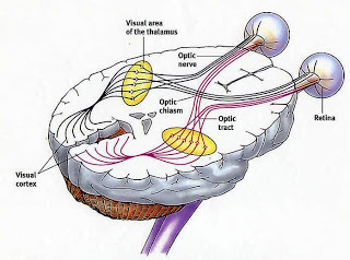
The relationship between the brain and the retinas
|
|
http://www.sneretina.com/images/pic-eye.jpg
http://biology.about.com/od/humananatomybiology/a/anatomybrain.htm
http://linsenbardt.net/wp-content/uploads/2010/11/brain.jpg
https://curemagazine.s3.amazonaws.com/npi/Media/CDR0000686379.jpg
http://www.glaucoma.org/glaucoma/eye-brain%20illustration.jpg
http://homepage.psy.utexas.edu/homepage/class/Psy308/Salinas/Vision/10.gif
http://3.bp.blogspot.com/_pGhYguBB1Sg/SMVEqK2XGXI/AAAAAAAAAI4/K1Sk1ZpStls/s320/Visual_pathway.jpg
http://biology-forums.com/gallery/14755_27_08_12_12_24_59_81982202.jpeg
http://astronomer.wpengine.netdna-cdn.com/wp-content/uploads/2008/03/rods_cones.gif
http://www.ib.bioninja.com.au/_Media/retina.jpeg
